


Home / Training / Manuals / Atlas of breast cancer early detection / Cases
Atlas of breast cancer early detection
Go back to the list of case studies
.png) Click on the pictures to magnify and display the legends
Click on the pictures to magnify and display the legends
BI-RADS Category (Left BCS, post operative breast): 2 (benign)
| Case number: | 163 |
| Age: | 51 |
| Clinical presentation: | Perimenopausal woman with average risk of breast cancer presented with left mastalgia. Clinical breast examination did not reveal any abnormality. She was diagnosed with left breast cancer and underwent surgery. At postoperative follow-up surveillance, she presented with a left breast lump and pain at the site of surgery. On examination, a firm palpable lump was detected beneath the surgical scar. |
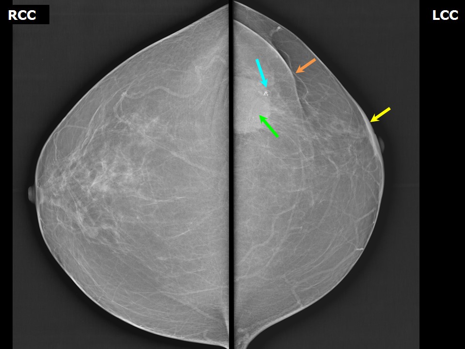
Mammography:
 |  |
 |
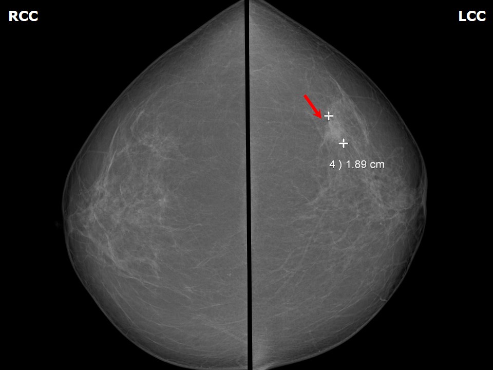
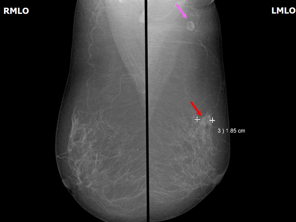
| Breast composition: | ACR category b (there are scattered areas of fibroglandular density) | Mammography features: |
| ‣ Location of the lesion: | 2014: Left breast, upper outer quadrant at 12 oclock, middle third |
| ‣ Mass: | |
| • Number: | 1 |
| • Size: | 1.8 cm in greatest dimension |
| • Shape: | Irregular |
| • Margins: | Indistinct |
| • Density: | High |
| ‣ Calcifications: | |
| • Typically benign: | None |
| • Suspicious: | None |
| • Distribution: | None |
| ‣ Architectural distortion: | Present |
| ‣ Asymmetry: | None |
| ‣ Intramammary node: | None |
| ‣ Skin lesion: | None |
| ‣ Solitary dilated duct: | None |
| ‣ Associated features: | None |
 | 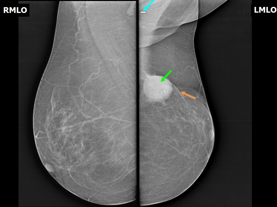 |
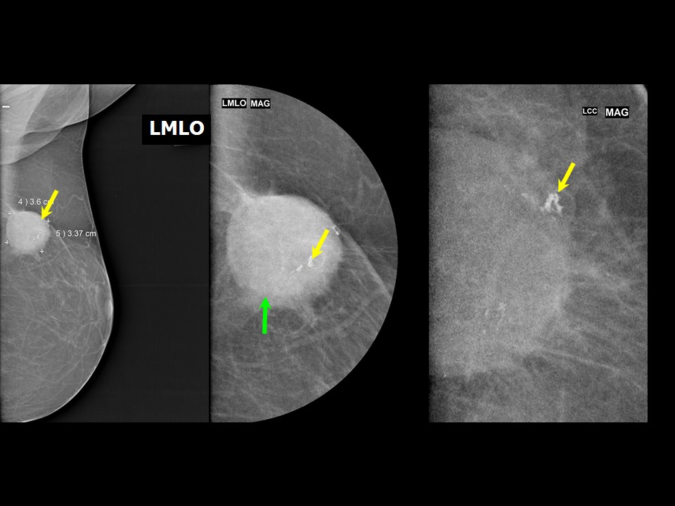 |
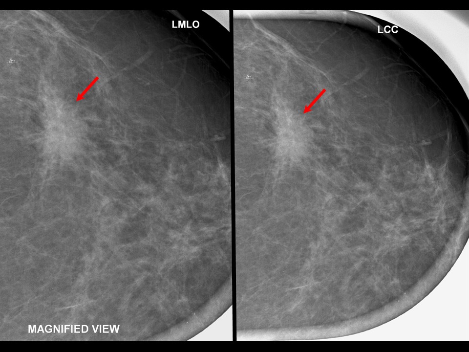
| Breast composition: | ACR category b (there are scattered areas of fibroglandular density) | Mammography features: |
| ‣ Location of the lesion: | 2020: Left breast, upper outer quadrant at 12 oclock, posterior third, beneath surgical scar |
| ‣ Mass: | |
| • Number: | 1 |
| • Size: | 3.8 cm |
| • Shape: | Round |
| • Margins: | Circumscribed |
| • Density: | High |
| ‣ Calcifications: | |
| • Typically benign: | Yes |
| • Suspicious: | None |
| • Distribution: | None |
| ‣ Architectural distortion: | None |
| ‣ Asymmetry: | None |
| ‣ Intramammary node: | None |
| ‣ Skin lesion: | None |
| ‣ Solitary dilated duct: | None |
| ‣ Associated features: | Dystrophic calcification within mass |
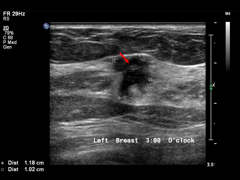
Ultrasound:
 |  |
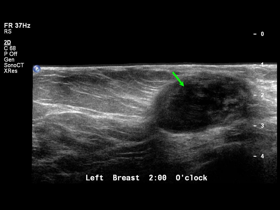
| Ultrasound features: 2020, 6 year postoperative surveillance: Left breast, upper outer quadrant at 2 oclock | |
| ‣ Mass | |
| • Location: | 2020, 6 year postoperative surveillance: Left breast, upper outer quadrant at 2 oclock |
| • Number: | 1 |
| • Size: | 1.8 cm |
| • Shape: | Irregular |
| • Orientation: | Not parallel |
| • Margins: | Angular |
| • Echo pattern: | Hypoechoic |
| • Posterior features: | No posterior features |
| ‣ Calcifications: | None |
| ‣ Associated features: | None |
| ‣ Special cases: | None |
 | 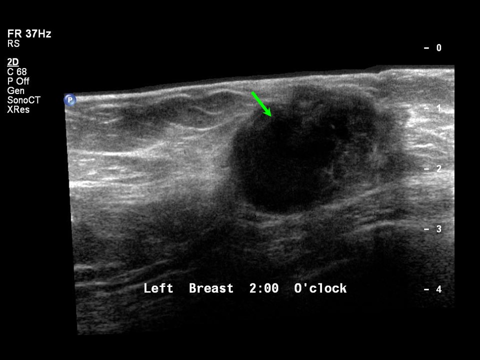 |
| Ultrasound features: Left breast, upper outer quadrant at 2 oclock | |
| ‣ Mass | |
| • Location: | Left breast, upper outer quadrant at 2 oclock |
| • Number: | 1 |
| • Size: | 3.0 × 2.5 cm |
| • Shape: | Oval |
| • Orientation: | Parallel |
| • Margins: | Circumscribed |
| • Echo pattern: | Heteroechoic with central anechoic component |
| • Posterior features: | No posterior features |
| ‣ Calcifications: | None |
| ‣ Associated features: | None |
| ‣ Special cases: | None |
BI-RADS:
BI-RADS Category: 4C (high suspicion for malignancy)BI-RADS Category (Left BCS, post operative breast): 2 (benign)
Further assessment:
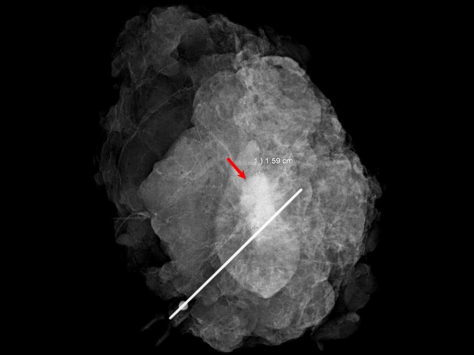
Further assessment advised: Referral for cytologyCytology:
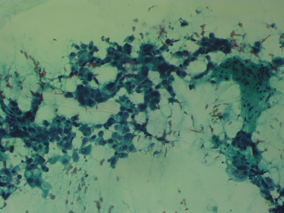 |
| Cytology features: | |
| ‣ Type of sample: | FNAC |
| ‣ Site of biopsy: | |
| • Laterality: | Left |
| • Quadrant: | Upper outer |
| • Localization technique: | Ultrasound-guided, no palpable lump |
| • Nature of aspirate: | Whitish |
| ‣ Cytological description: | Smears reveal numerous fragments of fibroadipose tissue with few loose clusters of malignant cells. Few scattered single isolated malignant cells are also seen |
| ‣ Reporting category: | Malignant |
| ‣ Diagnosis: | Carcinoma |
| ‣ Comments: | None |
Histopathology:
Breast-conserving surgery
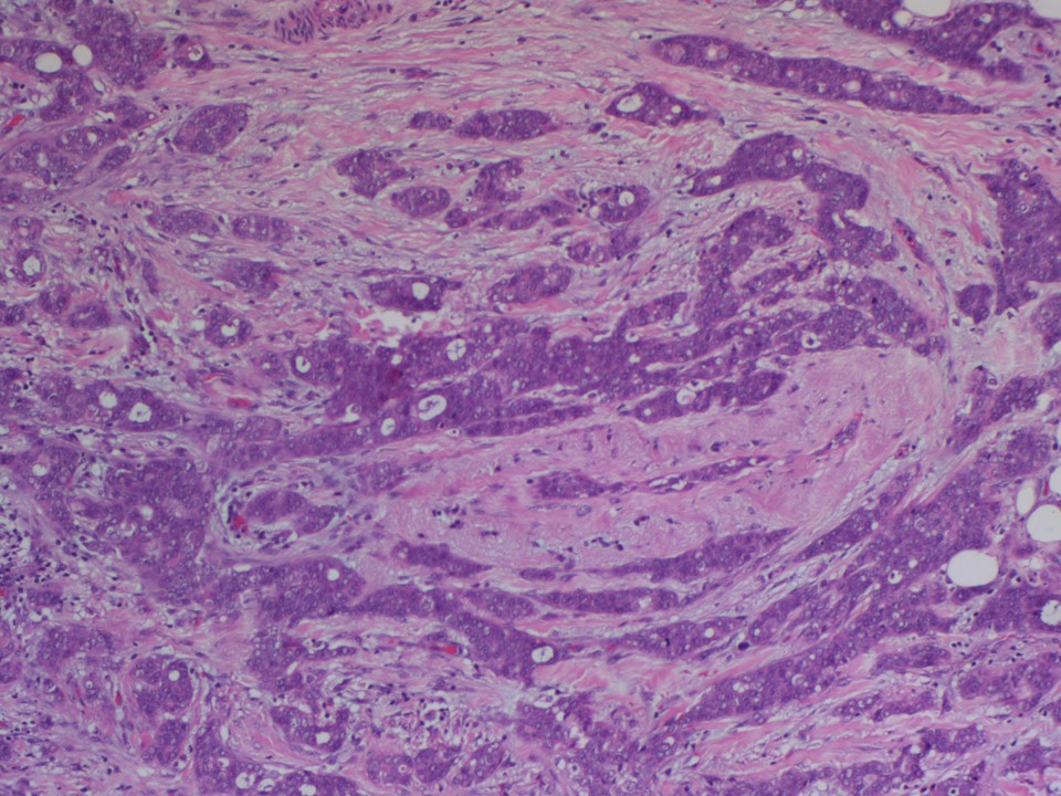 |
| Histopathology features: | |
| ‣ Specimen type: | Breast-conserving surgery |
| ‣ Laterality: | Left |
| ‣ Macroscopy: | Received a lumpectomy specimen oriented with long suture laterally and short suture superiorly. The specimen measured 6.0 × 5.0 × 4.0 cm. Skin flap measuring 2.7 × 2.8 cm. On serial sectioning an ill-defined whitish tumour with adjacent fibrosed areas identified measuring 1.7 × 1.4 × 1.0 cm. It is located 2.4 cm from the skin i.e. anterior margin, 1.6 cm from the posterior margin, 1.7 cm from the superior margin, 0.8 cm from the inferior margin, 1.1 cm from the medial margin, and 1.8 cm from the lateral margin. The remaining breast parenchyma is normal |
| ‣ Histological type: | Invasive breast carcinoma of no special type |
| ‣ Histological grade: | Grade 2 (2 + 3 + 1 = 6) |
| ‣ Mitosis: | 6 |
| ‣ Maximum invasive tumour size: | 1.7 |
| ‣ Lymph node status: | 0/20 |
| ‣ Peritumoural lymphovascular invasion: | Absent |
| ‣ DCIS/EIC: | DCIS - cribriform type, low grade |
| ‣ Margins: | Free of tumour |
| ‣ Pathological stage: | pT1cNo |
| ‣ Biomarkers: | ER positive; 100% of cells show nuclear staining with moderate intensity. Allred score 7/8 PR positive; 100% of cells show nuclear staining with moderate intensity. Allred score 7/8. HER 2 negative |
| ‣ Comments: | The adjacent breast shows fibrocystic change, apocrine and columnar metaplasia, areas of atypical ductal hyperplasia and usual ductal hyperplasia |
Case summary:
| Perimenopausal woman on postoperative surveillance; serial follow-up imaging showed stable seroma. No suspicious interval change was seen. An organized seroma with dystrophic calcification on the 6th year of follow-up mammogram is shown in this case. BI-RADS 2. |
Learning points:
|